Figure 3.

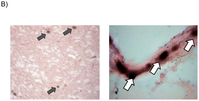
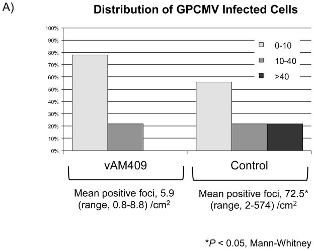
Reduced placental infection in vAM409-immunized dams compared to controls. (A) Distribution of in situ positive foci in vAM409-vaccinated (left panel) and control (right panel) animals. An average of 72.5 (range, 2–574) positive foci/cm2 were noted in control placentas. In contrast, in placentas of vAM409-vaccinated animals, an average of 5.9 positive foci (range, 0.8–8.8)/cm2 were noted (P < 0.05). (B) Demonstration of positive signal in control placenta. Left panel, 20× magnification of in situ labeled control dam demonstrating multiple positive foci (arrows) of GPCMV infection. Right panel, 80× magnification demonstrating positive cells (arrows) in trophoblasts of infected placenta.

