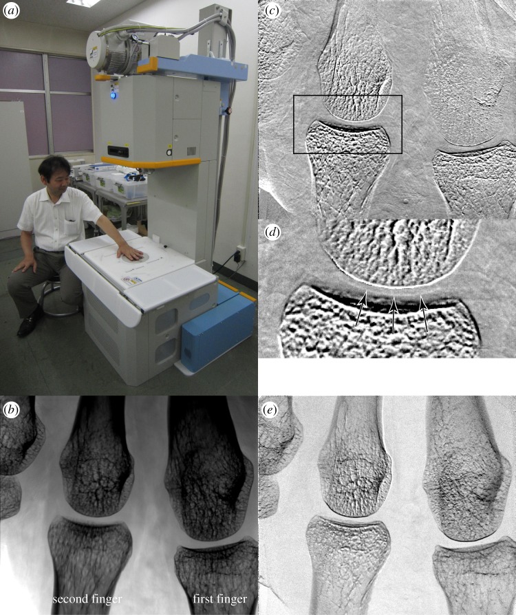
Figure 2.
(a) An X-ray Talbot–Lau phase imaging system installed in a hospital, and resultant (b) absorption image, (c) differential phase image, (d) its zoomed image at the rectangle indicated in (c), and (e) visibility image of a part of the first author's palm. X-ray refraction in the vertical direction of the images is sensed. Cartilage in a joint is revealed as indicated by the arrows in (d). The blurry feature in the upper right of (c) is considered to be due to movement during the measurement. (Online version in colour.)

