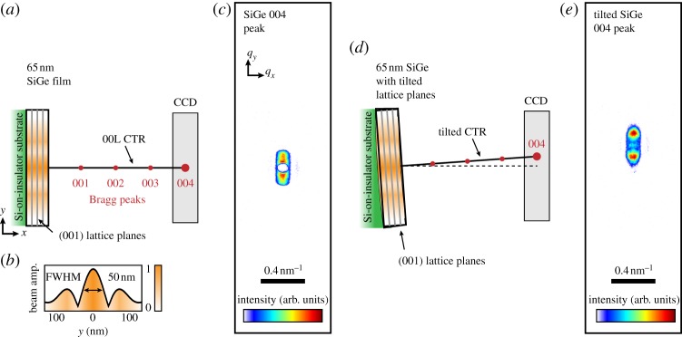
Figure3.

Schematics of a symmetric coherent nanodiffraction experiment in which two different regions of a 65 nm-thick epitaxial SiGe film grown on a silicon-on-insulator substrate were illuminated with nanofocused X-rays at the NPI are shown along with measured data. (a) A flat section of the film illuminated with the shaded beam profile shown in (b). (b) A calculation of an ideal focused X-ray profile at the NPI. In (a,d), the scattering plane is horizontal and normal to the page. The (001) lattice planes in the crystal films are depicted by the grey lines. The specular Bragg peak positions are depicted by circles along the crystal truncation rod (small circles indicate a forbidden SiGe reflection), and the measured 004 Bragg peak is shown in (c). A region of the film that was slightly tilted is depicted in (d) in which the 004 Bragg peak is displaced from the scattering plane, seen experimentally as a shift of the Bragg peak from the centre of the detector in (e). (Online version in colour.)
