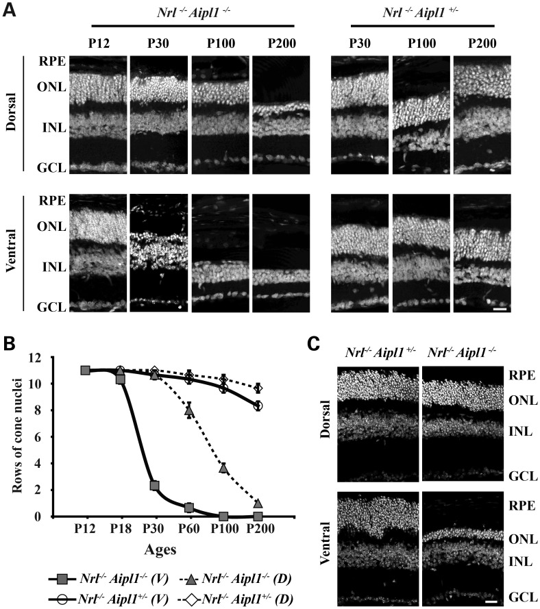
Figure 2.
Loss of Aipl1 leads to rapid and differential degeneration of cones in retina. (A) Retinal sections from all-cone mice lacking Aipl1 and littermate control stained with DAPI showing laminated nuclear layers. Top panel represents sections from the dorsal region of retina at various ages from P12 to P200. Sections from the ventral region of retina are shown in the bottom panel. (B) Quantitation of rows of outer nuclei in the dorsal (D) and ventral (V) regions of Nrl−/− Aipl1+/−and Nrl−/− Aipl1−/− at various ages. Counts were performed as described previously (32). Counts are an average from retinal sections at four different locations obtained from at least three sections each from three independent animals. (C) Retinal sections from all-cone mice lacking Aipl1 and littermate control reared in complete darkness. All sections were stained with DAPI showing laminated nuclear layers. Top panel represents sections from the dorsal region of retina at P30. Sections from the ventral region of retina are shown in the bottom panel. RPE, retinal pigment epithelium; ONL, outer nuclear layer; INL, inner nuclear layer and GCL, ganglion cell layer. Scale bar = 10 μm applies to all panels.

