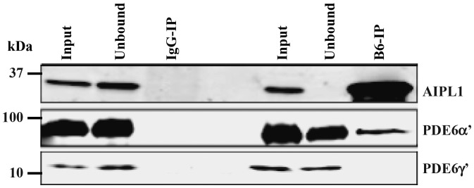
Figure 4.

Aipl1 interacts with catalytic subunit of cone PDE6. IP of Aipl1 using a mouse monoclonal antibody (B6-IP) from retinal extracts of all-cone mice (Nrl−/−) (right panel). IP with non-specific mouse IgG serves as a control (left panel). Aipl1, PDE6α′ and PDE6γ′ subunits were analyzed by immunoblotting. IP samples are five times more concentrated than input or soluble fraction. Protein molecular weight marker is shown on the left.
