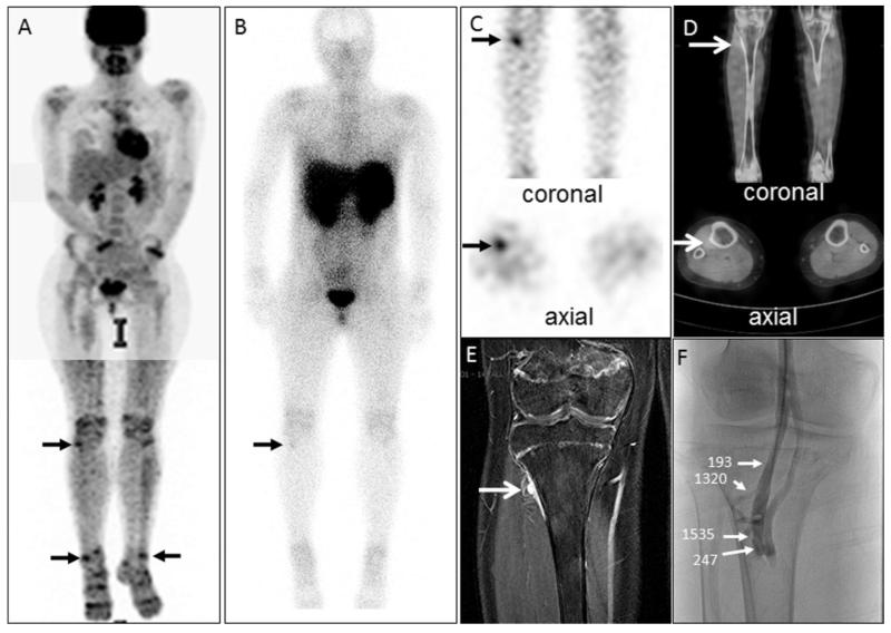
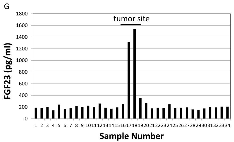
Figure 3.
An example of a case illustrating octreo-SPECT/CT and FDG-PET/CT complementarity: A 15-year-old girl who was found to have tumor-induced osteomalacia had an FDG-PET/CT study that demonstrated many areas of physiologic (e.g., brain, heart, kidneys) and non-physiologic (e.g., arrows) uptake (A). Subsequently an octreo-SPECT/CT study was performed that initially was read as negative (B). Guided by the non-physiologic FDG-PET/CT findings, a reevaluation of the octreo-SPECT/CT identified a suspicious lesion on the lateral aspect of the proximal right tibia on SPECT (C) and fused SPECT/CT (D) images (arrows). In retrospect the lesion was evident on the whole body image (B, arrow). Anatomic imaging identified a tumor on MRI (E). Further confirmation was deemed necessary prior to surgical resection, so venous sampling (VS) was performed. VS revealed a clear step-up in the region of the tumor suggested by functional and anatomical imaging (F&G). In panel F, the values indicate the intact FGF23 concentration at the location indicated by the arrows. In panel G, values from sites sampled are shown. The peripheral value was 188 pg/ml and the values at sites adjacent to the lesion were 1320 and 1535 pg/ml. The lesion was resected and the subject was cured.


