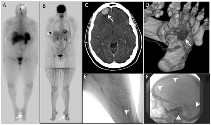
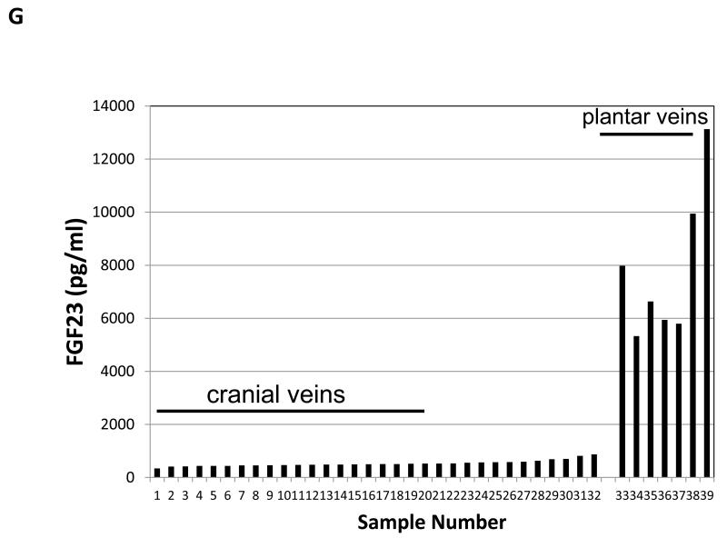
Figure 4.
An example of venous sampling in discriminating between two potential sites of tumor. Whole body octreo-SPECT was performed and identifed two suspicious lesions, white arrows (A). FDG-PET/CT suggested multiple lesions, including the lesion in the foot (B). The white asterisk (B) indicates the injection site. Anatomincal imaging studies confirmed the presence of a lesion at the brain-bone interface on CT scan (C), as well as a lesion on the plantar surface as shown on 3D CT scan (D). Venous sampling at these two sites was performed (E& F). In panels E & F, the course of the catheter the tips are indicated by white arrow heads. Measurement of intact FGF23 levels clearly revealed that the lesion in the foot was the source of the FGF23 (G).


