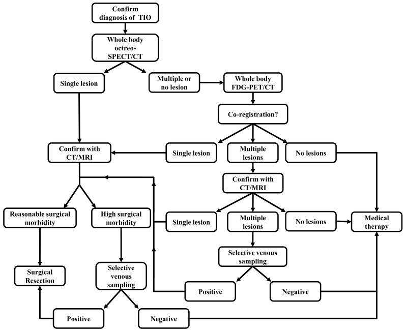
Figure 5.
Alogorithm for systematic approach to localizing tumors in tumor-induced osteomalacia. After confirming a clinical and biochemical picture of tumor-induced osteomalacia, we recommend obtaining functional imaging as a first step in tumor localization. Octreo-SPECT (SPECT/CT if available) is the recommended initial screening imaging test, as it was shown to have greater specificity and sensitivity than FDG-PET/CT. If a single lesion suspcious for TIO is found and it is confirmed on anatomical imaing and carries a reasonable surgical morbidity, surgical resection is recommended. If multiple lesions or no lesions are found on octreo-SPECT, FDG-PET/CT should follow. This may help to identify a lesion that was not initially seen on octreo-SPECT (as in Fig. 3), or help to confirm a lesion seen on octreo-SPECT. If appropriate, proceed to anatomical imaging with CT and/or MRI. If the area with the suspected lesion entails high surgical morbidity, or if multiple lesions are identified, selective venous sampling is recommended to confirm or rule out the tumor. If no lesion is found or a suspected lesion(s) is not confirmed on venous sampling, medical therapy is recommended.

