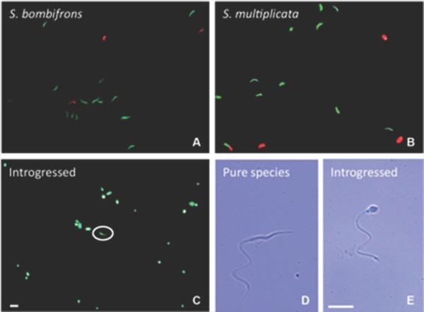
Figure 6.
Sperm viability and morphology. Epifluorescence images of live/dead staining of sperm (green: live cells; red: dead cells) in spermatic urine of S. bombifrons (A), S. multiplicata (B) and an introgressed individual (C). Typical (D) and abnormal (E) spermatozoid morphology are shown with bright field images at a higher magnification (400X). The white circle in (C) indicates a normally shaped spermatozoid (as in D) in the sample collected from the introgressed male. Both scale bars = 100 μm.

