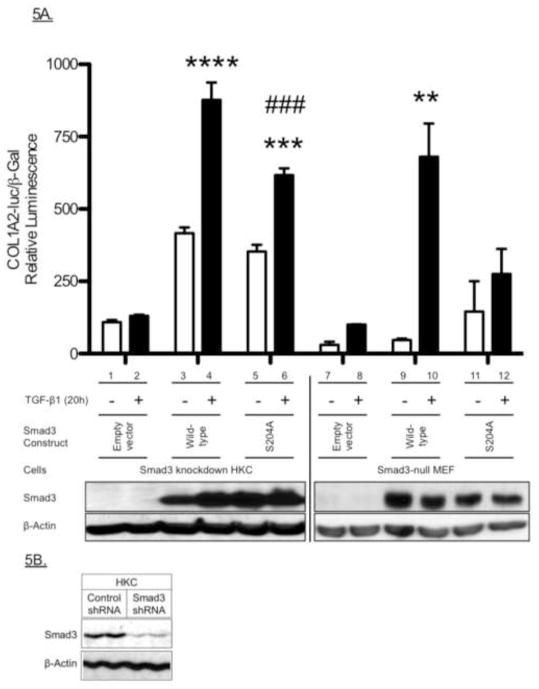
Figure 5. Role of the Serine-204 (S204) Smad3LR Phosphorylation Site in Smad3-mediated renal epithelial cell COL1A2 promoter activity.
A, Effect of the S204A-Smad3 mutant on Smad3-mediated COL1A2 promoter activity in renal epithelial cells. Smad3-knockdown HKC (bars 1–6) and Smad3-null MEFs (bars 7–12) were transfected with empty vector or pCS2-Smad3 constructs (wild-type or S204A), including the COL1A2-luciferase and β-galactosidase constructs for 3 hours. The transfected cells were then stimulated with TGF-β (2 ng/ml) for a further 20 h. Relative luciferase activity (mean ± S.E.M., n=3) was corrected for β-galactosidase activity. Shown here is a representative experiment of three independent experiments. *P<0.05, **P<0.01 vs. vehicle-treated cells. B, Smad3-knockdown in renal epithelial cells. Western blotting analysis of HKC stably expressing control- or Smad3-pGIPZ lentiviral shRNA.

