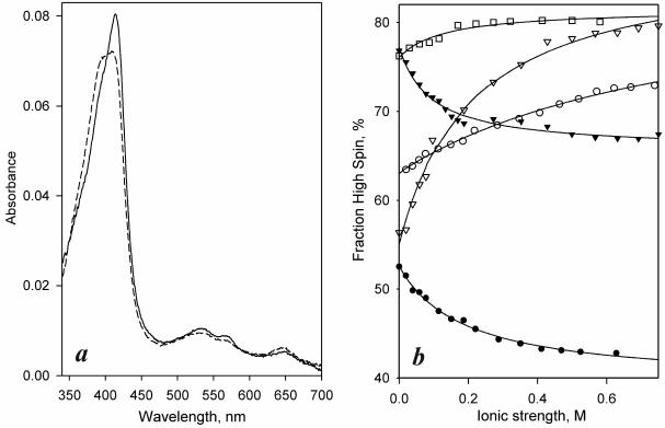
Figure 3.
Differences in position of spin equilibrium and their dependencies on ionic strength among different forms of CYP261 enzymes. (a) Absorbance spectra of purified CYP261D1− and CYP261D1(+) conformers (solid and dashed lines respectively). (b) Effect of ionic strength on the spin state of CYP261C2 (open squares) and the (+) (triangles) and − (circles) conformers of CYP261C1 (open symbols) and CYP261D1 (closed symbols). Solid lines represent the approximations of the data sets with a hyperbolic equation.

