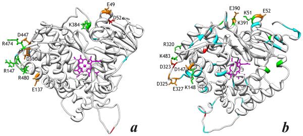
Figure 5.
Homology model of the structucture of piezophilic CYP261C1 (a) and CYP261D1 (b). The positions of the substitutions that result in appearance, elimination or substitution of charged residues (D,E,K,R) are highlighted in green (Arg and Lys residues) and red (Glu and Asp). Positions of other substitutions are shown in cyan. The substitutions presumably involved in the mechanism of high-pressure adaptation are highlighted by displaying their side chains. The side chains of potentially important Glu and Asp residues involved in charge-pairing interactions, which are conserved within each of the CYP261C1/C2 and CYP261D1/D2 pairs, are highlighted in orange.

