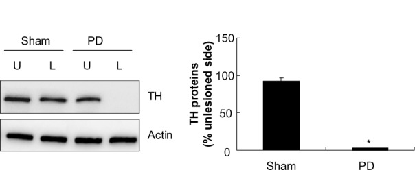
Figure 1.

Tyrosine hydroxylase levels in the striatum.
Notes: Western blot for TH of extracts from striatums of sham and 6-OHDA–lesioned rats. The optical density quantified by densitometry and the value of the lesioned side is expressed as percentage of the unlesioned striatum (lesioned/unlesioned × 100% ± SEM). *P<0.01 versus sham.
Abbreviations: PD, Parkinson’s disease; U, unlesioned side; L, lesioned side; TH, tyrosine hydroxylase; SEM, standard error of the mean.
