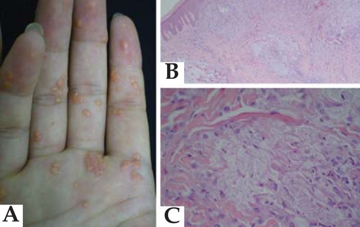
FIGURE 2.
A: Palmar region showing yellowish papules with erythematous halo. B: Rectified and epidermal inflammatory infiltrate composed of lymphocytes and xanthomized histiocytes in upper and middle dermis. (Hematoxylin–eosin; 40× magnification). C: Detail of xanthomized histiocytes (hematoxylin–eosin; 400× magnification)

