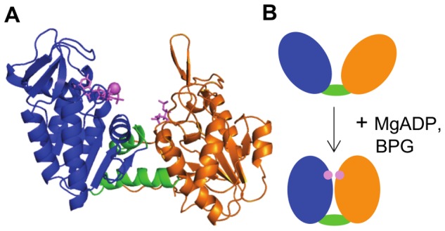
Figure 1. Structure of hPGK.

(A) Cartoon representation of X-ray structure of hPGK. N-domain, orange; C-domain, blue; interdomain region, green; substrates (BPG and ADP), purple sticks; Mg ion, purple sphere. (B) The hinge bending mechanism of hPGK. Coloring as in (A). Substrate binding triggers a closure of the two domains, thereby bringing the two reaction partners into close proximity.
