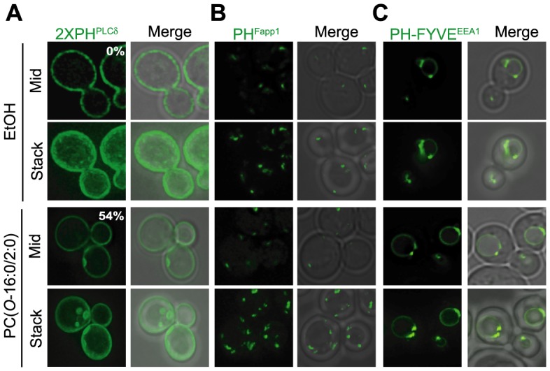
Figure 1. PtdIns(4,5)P2 is redistributed in response to PC(O-16:0/2:0).

Wild type (WT) cells (YPH500) expressing (A) GFP-2×PHPLCδ (PtdIns(4,5)P2) (B) GFP-PHFapp (PtdIns(4)P) or (C) GFP-FYVEEEA1 (PtdIns(3)P) were treated with either vehicle (EtOH) or PC(O-16:0/2:0) (20 µM, 15 min) and localization of the GFP probe quantified. The percentage of cells displaying a redistribution of the fluorescent reporter is reported in the inset of the figure.
