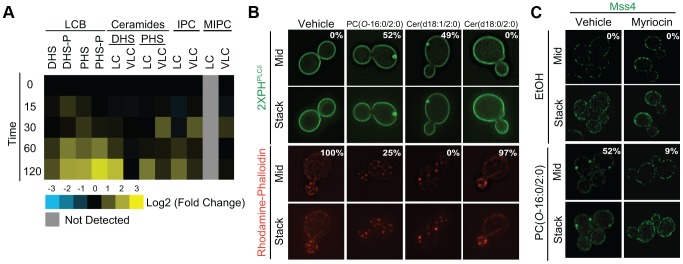
Figure 4. PC(O-16:0/2:0) disrupts sphingolipid metabolism leading to changes in Mss4-GFP localization.

(A) PC( O -16:0/2:0) treatment disrupts sphingolipid metabolism. Wild type (BY4741) cells were treated with vehicle or PC(O-16:0/2:0) (20 µM) for the indicated times (min). Lipids were extracted and sphingolipid levels were quantified and expressed as a log2 fold change of PC(O-16:0/2:0) treated from vehicle treated control. LCB, long chain base; IPC, inositol phosphorylceramide; MIPC, mannosyl phosphorylceramide; DHS(-P), dihydrosphingosine (1-phosphate); PHS(-P), phytohydrosphingosine; LC, long chain (acyl chain is equal to or less than 22 carbons); VLC, very long chain (more than 22 carbons). (B) Treatment with ceramide promotes PES formation and inhibits actin cytoskeleton polarization. Wild type (BY4741) cells expressing GFP-2×PHPLCδ were grown in YPD in the presence of vehicle (EtOH), PC(O-16:0/2:0), Cer(d18:1/2:0) or Cer(d18:0/2:0) (20 µM, 15 min) prior to imaging live or fixing and staining for acting cytoskeleton polarization as described in methods. The percentage of cells displaying a redistribution of the fluorescent reporter or proper actin polarization is reported in the inset of the respective figure. (C) Inhibition of sphingolipid metabolism prevents the relocalization of Mss4. Mss4-GFP (YKB2955) expressing cells were pretreated with vehicle or myriocin (5 µM) for 30 min and subsequently treated with vehicle or PC(O-16:0/2:0) (20 µM, 15 min) as previously done. Pretreatment with myriocin inhibited PC(O-16:0/2:0)-dependent changes in PES formation.
