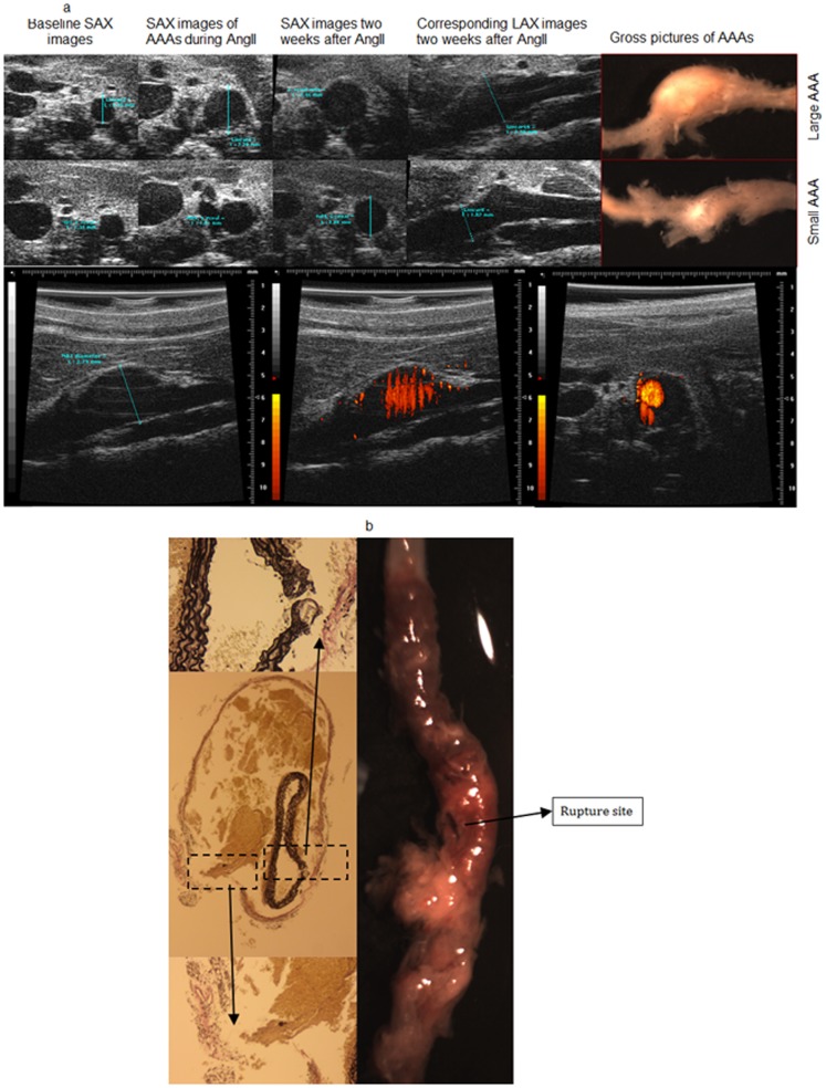
Figure 1. Representative images of AAA.
(A) Representative in vivo US and gross anatomic images of intact AAA. Top panels: Representative SAX and LAX images with corresponding gross anatomical pictures of a large and small AAA, respectively. Bottom: Representative US Power Doppler imaging technique employed for target-vessel confirmation. Power Doppler images delineate blood flow in a large AAA in LAX and SAX views. US, ultrasound; SAX, short axis; LAX, long axis; Ang II, angiotensin II; AAA, abdominal aortic aneurysm. (B): Representative image of gross anatomic and histological section of a ruptured AAA. Left: Middle panel is an elastin stained cross section demonstrating elastin and adventitial disruption of an AAA; the top and bottom panels are magnified areas of elastin and adventitial break, respectively. Right: Gross image of an abdominal aorta demonstrating a ruptured aneurysm. AAA, abdominal aortic aneurysm.

