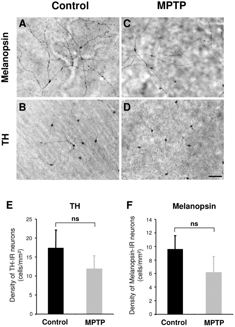
Figure 8. In the retina, the morphology of melanopsin and TH immunoreactive neurons appear normal after MPTP treatment.
The photomicrographs are from representative flat-mounted retina immunostained for melanopsin (A, C) or TH (B, D) in control (A, B) and MPTP-treated animals (C, D). Scale bar: 100 µm. E and F illustrate histograms of the densities of TH and melanopsin immunoreactive neurons. (Controls n = 2; MPTP n = 3).

