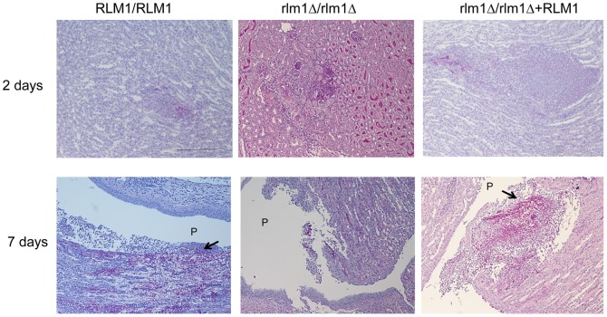
Figure 7. Kidney histology.
Representative photomicrographs of HE/PAS-stained paraffin sections of kidneys recovered from BALB/C mice infected with 5×10⁵ cells of wild-type SC5314 (RLM1/RLM1), mutant (Δrlm1/Δrlm1), and complemented SCRLM1K2A (Δrlm1/Δrlm1+ RLM1) C. albicans strains at 2 and 7 days post-i.v. infection. Arrows show hyphae invading the pelvis region. P-renal pelvis. Magnification of photographs: 100×. Bar: 100 µm for all photos.

