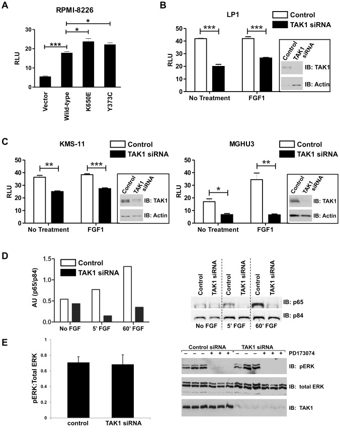
Figure 4. TAK1 knockdown inhibits FGFR3-dependent NFκB activation.
(A) 8226 (FGFR3 negative) MM cells were transfected with 5 µg FGFR3 constructs or empty vector, and NF-κB-Luc and pRL-TK control Renilla reporter at a ratio of 3∶1, respectively for 48 hours. Cells were then lysed and assayed for dual-luciferase activity. (B, C) FGFR3-expressing bladder and MM cell lines were transfected with control or TAK1 siRNA, and 24 hours later with κB-Luc and pRL-TK control Renilla reporter at a ratio of 3∶1. The following day, cells were serum-starved overnight and treated with ligand (FGF1) for 8 hours prior to lysis and dual-luciferase assay. (D) MGHU3 cells were transfected with TAK1 or non-targeting siRNA for 48 hours, serum starved overnight then treated with FGF1 ligand for the time indicated. Cells were then fractionated, and 10 µg of nuclear fraction was run on an SDS-page gel and western blotted. Blots were probed with anti-p65 and anti-p84 (nuclear marker) antibodies. Densitometry was performed and p65 measurements were normalized to p84 measurements. (E) FGFR3 signaling is not altered by TAK1 knockdown. KMS11 cells were transfected with control or TAK1 siRNA and, 24 hours later, treated with or without FGFR inhibitor, PD173074 for an additional 24 hours. Western blots were probed with p-ERK, total ERK and TAK1 antibodies. Statistical analysis was performed using a t-test; (*) p<0.05; (**) p<0.01; (***) p<0.001. Four independent experiments were performed.

