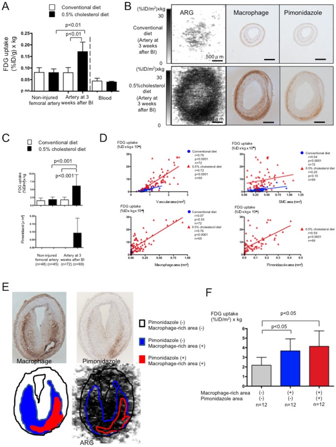
Figure 3. Arterial 18F-FDG uptake and its relationship to hypoxia.
A. Radioactivity levels in iliac-femoral arteries and blood. White and black bars, conventional and 0.5% cholesterol diets, respectively; n = 5). BI, balloon injury. B. Representative findings of macrophages and pimonidazole in the iliac-femoral arteries at three weeks after balloon injury in rabbits fed with conventional (upper row) or 0.5% cholesterol diet (lower row). Autoradiographic images show increased 18F-FDG uptake in the artery with macrophage-rich neointima. Immunohistochemical staining shows hypoxic area (pimonidazole positive area) localized in macrophage-rich area in deep portion of wall. C. Uptake of 18F-FDG and immunopositive area for pimonidazole in arterial sections. White and black bars, conventional and 0.5% cholesterol diets, respectively. D. Correlations between 18F-FDG uptake and vascular area, SMC-, macrophage-, and pimonidazole-immunopositive areas in sections of arteries at three weeks after BI. E. Representative trace of macrophage-rich and/or pimonidazole immunopositive area in autoradiographic image. Areas that were rich in macrophages or pimonidazole were traced on immunohistochemical images, and then FDG uptake was measured in corresponding areas of autoradiographic images. F. Autoradiogram of arteries with macrophage-rich neointima shows 18F-FDG uptake in areas with or without macrophage-rich area or pimonidazole immunopositivity.

