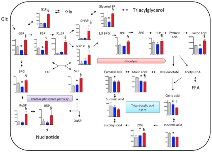
Figure 5. Levels of metabolites of glycolysis, the pentose phosphate pathway, tricarboxylic acid cycle and glyconeogenesis/glycogenolysis in cultured macrophages.
For M1 polarization, THP-1 cells were treated with PMA (320 nM) for 6 hours and then cultured with PMA plus LPS (10 ng/ml) and INFγ (20 ng/ml,) for another 42 hours. After replacement of culture medium, PMA-treated control macrophages (gray bar, n = 6) and M1 macrophages (blue bar, n = 6) were incubated under normoxic (21% O2) condition for 6 hours, or the M1 macrophages were incubated hypoxic (1% O2) conditions for 6 hours (red bar, n = 6). Metabolite levels are expressed as pmol/106 cell. *p<0.01 vs. PMA control, †p<0.05 vs. PMA control, §p<0.01 vs. M1 normoxia, ¶p<0.05 vs. M1 normoxia. 1,3BPG, 1,3-bisphosphoglycerate; 2PG, 2-phosphoglyceric acid; 3PG, 3-phosphoglyceric acid; 2OG, 2-oxoglutaric acid; 6PG, 6-phosphogluconic acid; DHAP, dihydroxyacetone phosphate; E4P, erythrose 4-phosphate; F1-6P, fructose 1,6-diphosphate; F6P, fructose 6-phosphate; FFA, free fatty acid; G1P, glucose 1-phosphate; G3P, glyceraldehyde 3-phosphate; G6P, glucose 6-phosphate; Glu, glucose; Gly, glycogen; PEP, phosphoenolpyruvic acid; R5P, ribose 5-phosphate; Ru5P, ribulose 5-phosphate; S7P, sedoheptulose 7-phosphate; Xu5P, xylulose 5-phosphate.

