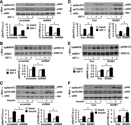
Figure 3.
SH2B1 promotes the PI 3-kinase/Akt pathway in β-cells. A–D: INS-1 832/13 cells were stably infected with scramble or shRNA vectors as described for Fig. 2A. A–C: Cells were deprived of serum overnight and stimulated with 50 nmol/L IGF-1 (A and B) or 100 nmol/L insulin (C) for 15 min. Cell extracts were immunoblotted with the indicated antibodies. Akt phosphorylation was quantified and normalized to total Akt levels, whereas phosphorylation of ERK1/2 was normalized to total ERK1/2 levels. Basal: n = 4–6; IGF-1: n = 4–6; insulin: n = 4. D–F: SH2B1β was stably overexpressed in INS-1 832/13 cells as described for Fig. 2D. Cells were stimulated with 50 nmol/L IGF-1 (D and E) or 100 nmol/L insulin (F) for 15 min, and Akt and ERK1/2 phosphorylation was measured as described above. Basal: n = 4; IGF-1: n = 4; insulin: n = 4; 2.8 mmol/L: n = 4–8. Data are means ± SE. *P < 0.05. a.u., arbitrary units; Con, control.

