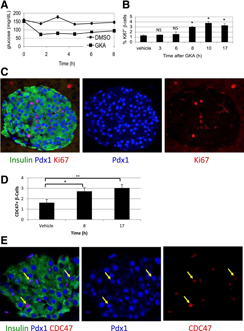
Figure 1.
Using GKA to define the time from quiescence to G1 in pancreatic β-cells in vivo. A: Blood glucose levels drop upon treatment of mice with a GKA, reflecting increased glycolysis-stimulated insulin secretion in β-cells. B: GKA increases the fraction of replicating β-cells 8 h after administration. C: Representative immunofluorescence image documenting replication of β-cells in vivo using the cell-cycle marker Ki67, 17 h after administration of GKA. D: The fraction of Cdc47+ β-cells increases 8 h after administration of GKA. E: Representative immunofluorescence image of Cdc47 staining in islets, 17 h after administration of GKA. Arrows point to Insulin+Pdx1+Cdc47+ cells. *P < 0.05, **P < 0.01. NS, not significant.

