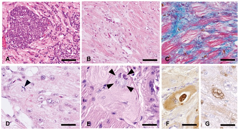
Figure 1. Histopathology and immunohistochemistry of hearts in Japanese native fowls.
(A) Mononuclear cells infiltrate into myocardial fibers. Chicken No. 5. Bar = 40 µm. (B) Disarrangement of myocardial fibers with interstitial fibrosis. Chicken No. 9. Bar = 90 µm. (C) Interstitial myocardial fibrosis confirmed by Masson's trichrome stain. Chicken No. 14. Bar = 25 µm. (D) Hypertrophied cardiomyocytes with multiple atypical nuclei. Mitosis (arrowhead) is occasionally noted in these cardiomyocytes. Chicken No. 14. Bar = 20 µm. (E) Matrix inclusion bodies (arrowhead) in the cytoplasm of Purkinje fibers. Chicken No. 14. Bar = 20 µm. (F) Purkinje fibers and matrix inclusion bodies positive for ALV common antigen. Chicken No. 14. Bar = 20 µm. (G) A large atypical nucleus in a hypertrophied cardiomyocyte is positive for PCNA. Chicken No. 14. Bar = 20 µm.

