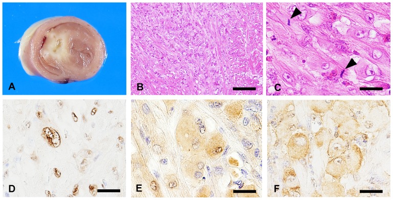
Figure 4. Gross pathology, histopathology and immunohistochemistry of chickens experimentally inoculated with Km_5666.
(A) Grayish-white ill-defined tissue was located in the septum at 35 days old. (B) Locally extensive growth of hypertrophied cardiomyocytes. Bar = 75 µm. (C) Mitosis (arrowhead) observed in hypertrophied cardiomyocytes. Bar = 30 µm. (D) Numerous nuclei in atypical myocardium were positive for PCNA. Bar = 20 µm. (E–F) Hypertrophied cardiomyocytes positive for p-Akt (E) and p-tuberin (F). Bars = 20 µm.

