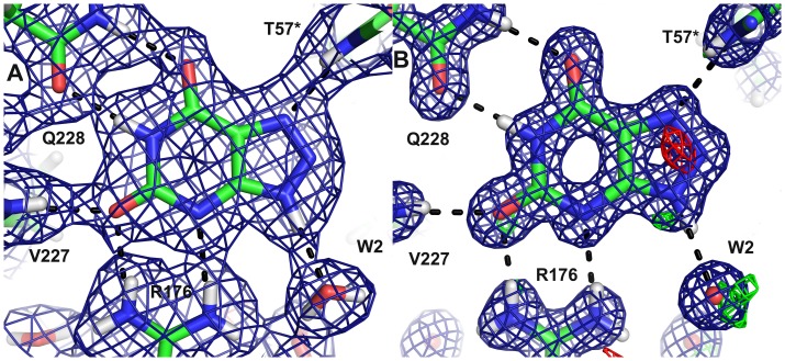
Figure 10. The active site with the inhibitor 8-azaxanthine in the neutron structure showing the 2mFo–DFc map contoured at 1.0 σ (blue) and the mFo–DFc map contoured at 3.0 σ (positive green, negative red).
B) The corresponding maps in the 1.1 Å X-ray structure contoured at 1.5 σ and 3.0 σ, respectively.

