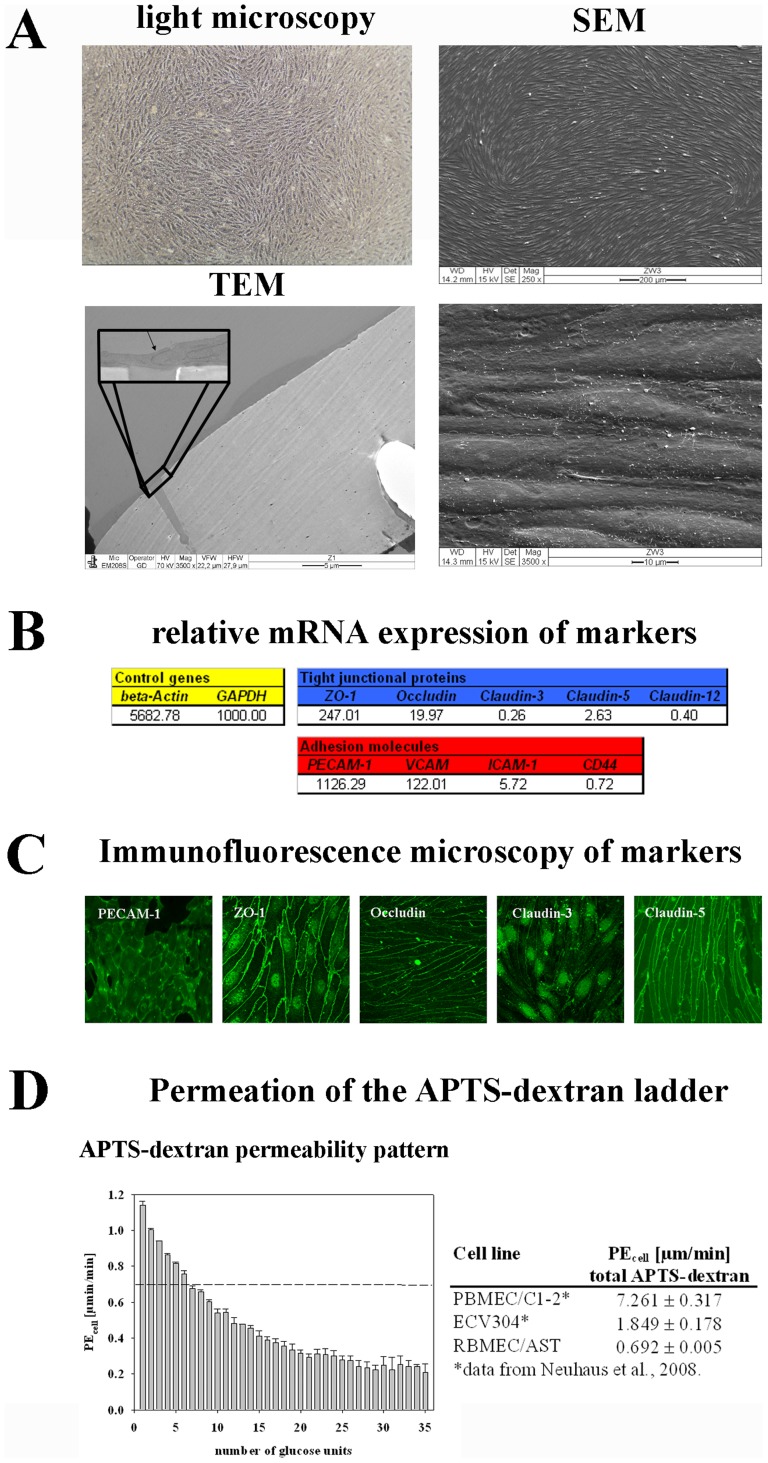
Figure 4. Characterization of the BBB model based on primary rat brain microvascular endothelial cells (RBMEC) and astrocytes.
RBMECs grow in endothelial cell typical spindle-like morphology proven by light and scanning electron microscopy (SEM). Transmission electron microscopic (TEM) images confirmed that RBMEC grow as a monolayer. The enlarged part of the image shows two RBMECs connected to each other directly over a pore of the Transwell insert membrane (A). mRNA expressions of tight junction proteins ZO-1, occludin, claudin-3, claudin-5 and claudin-12, and of adhesion molecules PECAM-1, VCAM, ICAM-1 and CD44. All data were related to endogenous control GAPDH which was set to 1000 (B). Immunofluorescence images of PECAM-1, ZO-1, occludin, claudin-3 and claudin-5 confirmed the protein’s presence and localization in RBMEC layers (C). Transport studies with paracellular marker APTS-dextran ladder confirmed functionality of the barrier. RBMEC layers were able to differentiate between the different dextran fractions in a molecular size-dependent manner. Comparison of the permeability coefficients for APTS-dextran across PBMEC/C1-2, ECV304 and RBMEC layers is presented in the table on the right side (D).

