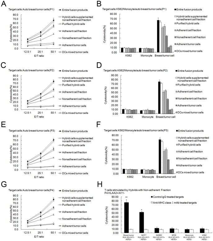
Figure 4. CTL assays induced by purified DC-tumor hybrids or other components of the fusion products.
The cytotoxicity assays were performed using CytoTox 96 Non-Radioactive Cytotoxicity Assay kit. (A, C, E, G) Non-adherent PBMCs stimulated with total fusion products (▪), hybrid cells supplemented with the non-adherent cell fraction (◊), purified hybrid cells (○), the adherent cell fraction (▴), the non-adherent cell fraction (△), adherent tumor cells (♦), or DCs mixed with tumor cells (□) for 7 days in the presence of 20 units/mL human IL-2 were used as the effector T cells. Then breast tumor cells were co-cultured with the effector T cells for 4 h at ratios of 1∶12.5, 1∶25, and 1∶50, respectively. Points, mean values of triplicate samples; bars, SD. The results showed that T lymphocytes activated by purified hybrids, the adherent cell fraction, the non-adherent cell fraction, total fusion products, purified hybrid cells supplemented with the non-adherent cell fraction lysed auto breast tumor cells much more effectively than T cells activated by adherent tumor cells or DC mixed with tumor cells (P<0.05). Lysis induced by total fusion products or purified hybrid cells supplemented with the non-adherent cell fraction was the most effective. Purified hybrid cells or the adherent cell fraction induced more effective lysis than the non-adherent cell fraction (P<0.05). There was no difference between total fusion products and purified hybrid cells supplemented with the non-adherent cell fraction or between purified hybrids and the adherent cell fraction (P>0.05). (B, D, F) Natural killer-sensitive K562 cells and monocytes were used as control targets in a parallel CTL assay, and the ratio for effector and target cells was 50∶1. Columns, mean values of triplicate samples; bars, SD. No lysis against K562 or monocytes was induced. H, T cells were stimulated by purified hybrid cells supplemented with the non-adherent cell fraction and then normal breast tumor cells lines MCF7, SKBR3 and BT20 were included as targets and MHC Class I molecule blocking test was done. Effector T can not only lyse the auto breast tumor cells (HLA A2+/A11−, HER2+), but also lyse the HLA-A2 matched MCF7 (HLA A2+/A11−, HER2+) to a less extent ( P<0.05) which can be blocked by preincubation with anti-MHC I antibody. (A B from patient1, C D from patient2, E F from patient3, G H from patient4).

