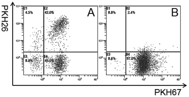
Figure 6. FACS analysis of apoptotic/necrosis tumor cells phagocytosed by DCs.

DCs and auto breast tumor cells (from patient 1) were stained red and green by PKH26 and PKH67 and double positive cells were analyzed using FACS and confocal microscopy. A, FACS analysis of the non-adherent cells fraction from the total fusion products. DC phagocytosis of apoptotic tumor cells was calculated as the percentage of double-positive cells, and was approximately 42%. B, FACS analysis of DCs mixed with tumor cells. No significant phagocytosis was observed.
