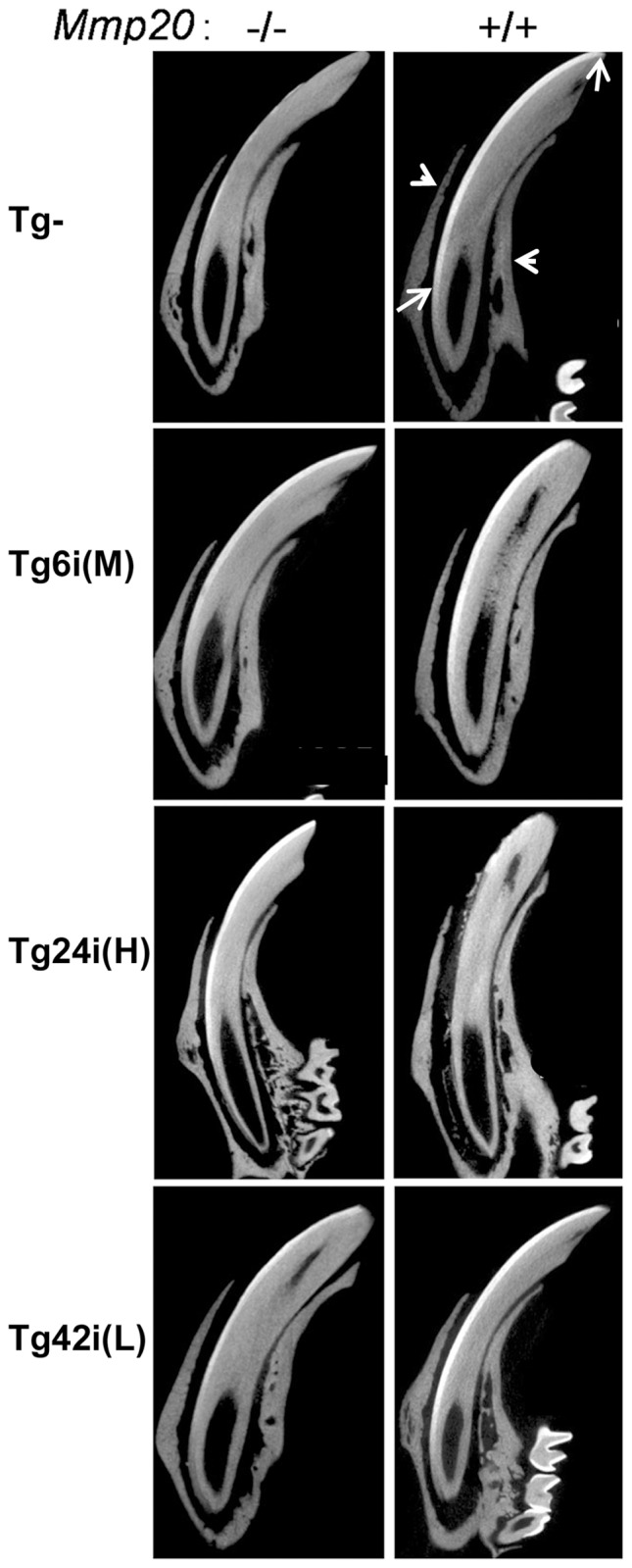
Figure 4. µCT analyses of incisor enamel from Mmp20 transgenics and controls.

Enamel on rodent incisors is present only on the labial side (arrows). The presented longitudinally oriented incisors were reconstructed from µCT images. The incisors protrude from bone (arrowheads) and are arranged so that the labial side is to the left. The wild-type incisors (Tg– MMP20+/+, top right panel) have a bright line of mineralized enamel that extends from the apical region (bottom arrow) to the labial incisal tip (top arrow). This mineralized enamel was mostly missing from the Mmp20 null incisors (Tg– Mmp20−/−, top left panel). The transgenic incisors in the null background (Tg+ Mmp20−/−) recovered some or most of the enamel layer along the labial surface. When the transgenes were present in the wild-type background (Tg+ Mmp20+/+), the enamel layer seemed relatively normal on incisors from mice transgenic for Tg6i (M) or Tg42i (L), but it was severely disrupted on the Tg24i (H) Mmp20+/+ incisors. For incisors, Tg24 was the highest, Tg6 the middle and Tg42 the lowest expressing transgene.
