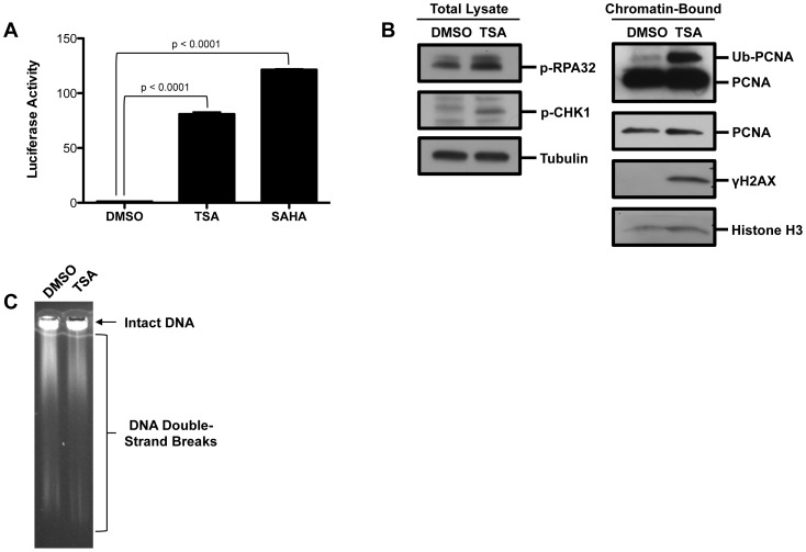
Figure 1. Trichostatin A induces DNA damage.
(A) HEK293T ELG1-LUC cells were treated with 0.75 µM TSA or 50 µM SAHA for 48 hr. LUC activity was measured using One-Glo. The graph represents average LUC activity ± SD. Significance was calculated using a two-tailed t-test. (B) HEK293T cells were treated with 50 µM TSA for 24 hr. Using the indicated antibodies proteins levels were visualized by Western blot analysis from either the chromatin-bound fraction or total cell lysate. (C) HEK293T cells were treated with 100 uM TSA for 24 hours and then Pulsed-field gel electrophoresis was performed.

