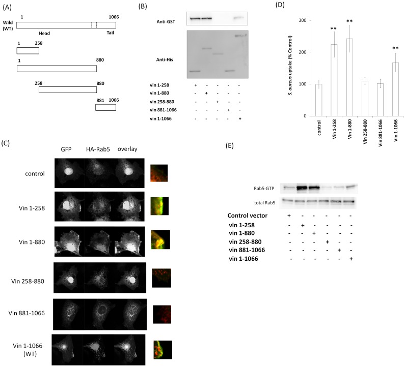
Figure 7. Determination of the Rab5-binding domain of vinculin.
(A) Schematic of vinculin deletion mutants. (B) GST-Rab5 (Q79L) was incubated with purified His-vinculin deletion mutants, and a His-pull-down assay was performed. The beads were assayed by western blotting. (C) GFP, GFP-Vin1-258, GFP-Vin1-880, GFP-Vin258-880, GFP-Vin881-1066 and GFP-vinculin (full length) were coexpressed with HA-Rab5 in Cos-7 cells and immunostained with anti-HA antibody. (D) pHrodo red-labeled S. aureus was added to the medium of Cos-7 cells expressing each vinculin deletion mutant and incubated for 2 h at 37°C. The graph shows mean ± S.E. values of six independent experiments, **p<0.01. (E) Cos-7 cells were transfected with HA-Rab5 (WT) and vinculin deletion mutants. S. aureus was added to the medium of transfected cells and incubated for 60 min at 37°C. The cells were lysed and subjected to a GST-R5BD pull-down assay. GST-R5BD-bound beads and lysates were assayed by western blotting with anti-HA antibody.

