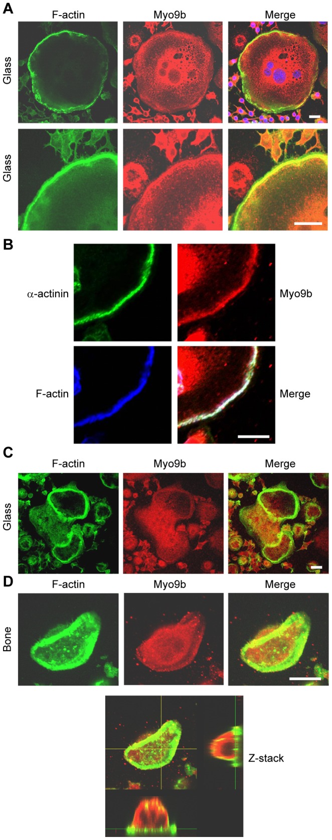
Figure 1. Distribution of Myo9b in osteoclasts.

Osteoclast podosomes and sealing zones were labeled with fluorescent phalloidin and anti-Myo9b antibodies and viewed by confocal microscopy. A, In mature osteoclasts on glass coverslips, Myo9b is at its highest levels in the perinuclear region and in the peripheral podosome belt. Nuclei are visible in blue in the merged image. B, In osteoclasts on glass coverslips, Myo9b associates closely with the podosome core protein α-actinin. C, In immature osteoclasts on glass that have not formed peripheral podosome belts, Myo9b is present, though not enriched, in internal podosome rings. D, Myo9b is mostly absent from sealing zones in osteoclasts on bone. A Z-stack image of an osteoclast on bone demonstrates that Myo9b is present throughout the cytoplasm. Scale bars = 20 µm.
