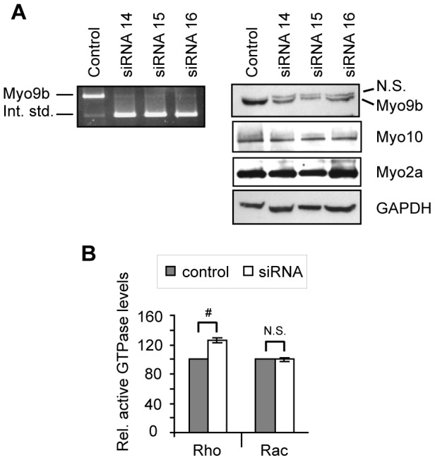
Figure 2. Knockdown of Myo9b increases cellular Rho activity.

A, Competitive RT-PCR (left panel) and Western blot (right panels) show efficient knockdown of Myo9b in mouse bone marrow-derived osteoclasts. For competitive RT-PCR, 1 pg of a synthetic RNA containing the Myo9b primer binding sites (the internal standard) was added to 1 µg sample total RNA, as described in Materials and Methods. RT-PCR then resulted in amplification of both cellular Myo9b and the internal standard, which served as a control for relative Myo9b mRNA levels. By Western analysis, knockdown of Myo9b did not significantly change protein expression of two other osteoclast myosins. GAPDH is shown as a loading control. N.S. = non-specific band. B, Protein pull-down followed by Western analysis and densitometry was used to quantify levels of active Rho or Rac in control or Myo9b siRNA-treated marrow-derived osteoclasts. Levels of active small G-proteins were normalized to levels of total Rho or Rac. The graph was compiled from three such experiments and shows that knockdown of Myo9b resulted in increased cellular levels of Rho but not Rac. #: P<0.001; N.S.: not significant.
