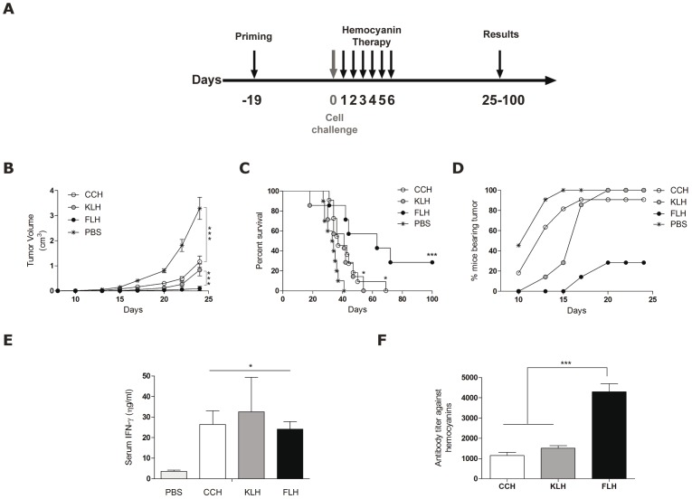
Figure 4. FLH possesses greater anti-tumor activity than CCH and KLH in the B16F10 mouse melanoma model.
(A). The schedule of the bioassay. Groups of 5–7 mice were sc primed with FLH, CCH or KLH (400 µg/100 µl PBS). After 19 days, the mice were challenged sc with 1.5×10⁵ B16F10 melanoma cells, and they then received intralesional injections with 100 µg of each hemocyanin for 6 consecutive days. Data are representative of two independent experiments. (B). Effect of FLH on melanoma growth. The tumor volume was measured every 3 days; ***p<0.001 between CCH, KLH and FLH versus PBS and ***p<0.001 between FLH versus CCH and KLH. (C). Animal survival. Kaplan-Meier survival analysis were compared using the log-rank test; ***p<0.001 FLH versus PBS, *p<0.05 between CCH and KLH versus PBS and *p<0.05 between CCH and KLH versus FLH. (D). Effect of FLH on melanoma growth. Tumor progression was measured for 25 days following the different treatments. (E). Levels of IFN-γ in the sera of mice. The levels of IFN-γ in mouse sera were determined by ELISA on day 13 of the bioassay; *p<0.05 hemocyanins versus the control with PBS. (F). Humoral immune response against hemocyanins. Anti-hemocyanin antibody titers in mouse sera were determined by ELISA on day 13 of the bioassay; ***p<0.001 FLH versus CCH and KLH.

