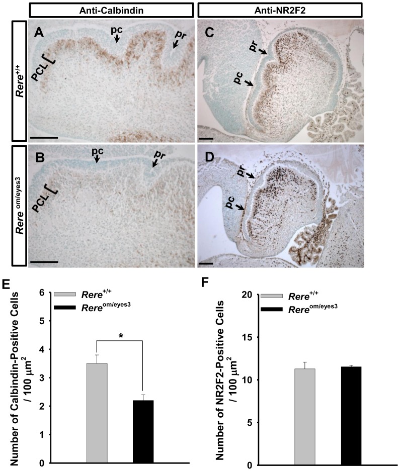
Figure 6. The migration and maturation of Purkinje cells was altered in Rere om/eyes3 embryos at E18.5.
A–D. Mid-sagittal sections of the vermis were probed with rabbit polyclonal anti-calbindin antibodies (A and B) or anti-NR2F2 antibodies (C and D). A–B. Number of calbindin-positive cells appeared to be lower in the cerebellums of Rere om/eyes3 embryos (B) when compared with those of their wild-type littermates (A). C–D. In the cerebellums of wild-type embryos, most of the NR2F2-positive cells were located in the Purkinje cell layer underneath the external granule cell layer at E18.5 (C). In contrast, the number of NR2F2-positve cells was increased in the center of cerebellar cortex of Rere om/eyes3 embryos at same time point (D). E-F. Calbindin-positive cells (E) and NR2F2-positive cells (F) were counted in the cerebellums of 18.5 embryos of both genotypes and normalized by the area of the cerebellum. The number of calbindin-positive cells/µm2 was significantly lower in the cerebellums of Rere om/eyes3 embryos when compared with wild-type littermates (* = p<0.03). However, number of NR2F2-positive cells/µm2 was comparable between Rere om/eyes3 embryos and their wild-type littermates. Quantification was performed using fifteen slides containing at least three sections from three independent littermates. pc, preculminate fissure; pr, primary fissure; PCL, Purkinje cell layer. Scale bar = 100 µm.

