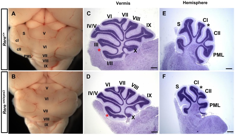
Figure 7. Rere om/eyes3 mice have cerebellar foliation defects at P21.
A–B. The cerebellar vermis and hemispheres were evident in wild-type and Rere om/eyes3 mice. C–D. Mid-sagittal sections of the vermis region were stained with cresyl violet. In the cerebellums of Rere om/eyes3 mice, the precentral fissure was severely attenuated and failed to divide lobule I/II from lobule III. Red asterisks indicate the location of the precentral fissure in wild-type mice (C) and marks the abnormal lobule in Rere om/eyes3 mice (D). E–F. Sagittal sections of the cerebellar hemispheres were stained with cresyl violet. The intercrural fissure was fully developed in wild-type mice (E), but was severely attenuated in the cerebellums of Rere om/eyes3 mice (F). The black asterisks indicate the intercrural fissure. Roman letters mark corresponding lobules in the vermis of the cerebellum. CI, lobule crus I; CII, lobule crus II; PML, paramedian lobe; S, simple lobule. Scale bar = 500 µm.

