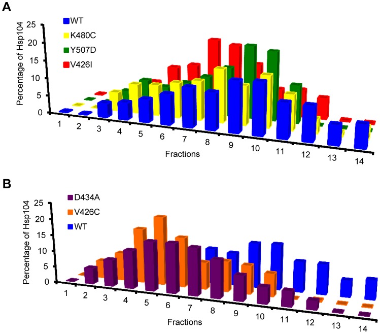
Figure 3. The M-domain plays a role in hexamer formation.
The oligomeric distribution of recombinant wild type (WT) Hsp104 (blue, A & B) and (A) Hsp104-V426I (red), Hsp104-K480C (yellow), and Hsp104-Y507D (green), or (B) Hsp104-V426C (orange) and Hsp104-D434A (purple), was analyzed by ultracentrifugation through a linear glycerol gradient in the presence of 5 mM ATP. Equal fractions from the gradients were collected and analyzed by western blot with an anti-Hsp104 antibody. The amount of Hsp104 in each fraction was quantified by ImageJ and graphed as a fraction of the total Hsp104. The gradients were repeated twice with recombinant protein from two separate recombinant protein purification preparations.

