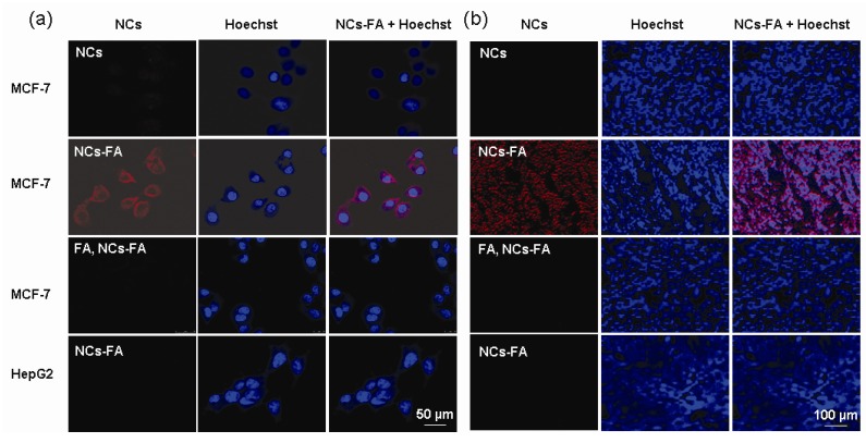
Figure 3.
The NCs-FA nanoprobes bind specifically. (a) MCF-7 and HepG2 cell were stained by the NCs-FA nanoprobes (NCs) and visualized by confocal laser microscopy, respectively. (b) A MCF-7 and HepG2 tumor tissue were stained by the NCs-FA nanoprobes (NCs) and visualized by fluorescent microscopy, respectively.

