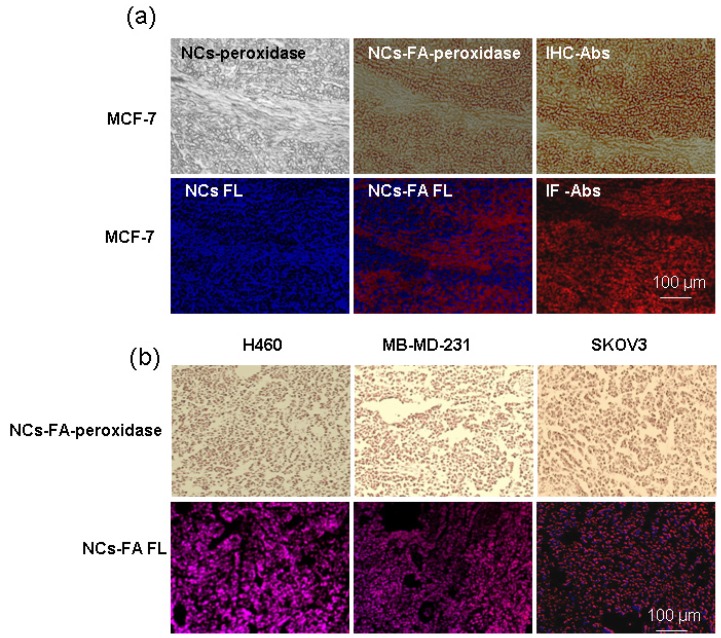
Figure 4.
The NCs-FA nanoprobes for histochemical stains. (a) MCF-7 tumor tissues were stained by the NCs (NCs-FA nanoprobes and Abs), and visualized by light microscopy and fluorescent (FL) microscopy, respectively. (b) H460, MDA-MB-231and SKOV3 tumor tissues were stained by the NCs-FA nanoprobes and visualized by light microscopy and fluorescent microscopy, respectively.

