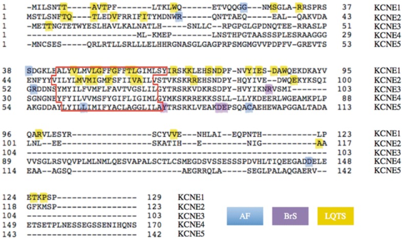
Figure 2.
Human KCNE1-KCNE5 protein sequence alignments and gene variants. Image of aligned sequences generated using http://www.uniprot.org/align. Colors highlight inherited or sporadic non-synonymous mutations or polymorphisms resulting in single amino acid changes (changes involving >1 amino acid are omitted). In cases where an amino acid substitution is associated with LQTS in addition to another arrhythmia, only the latter is color-coded (see Table S1 for full information). The predicted transmembrane domain for each subunit is outlined in red.

