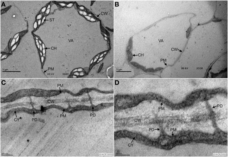
Figure 1.
Plasmodesmata are structures that persist under autophagy conditions. Three-week old Arabidopsis seedlings were exposed to 10 μ M fumonisin B1 for 4 days in order to induce programmed cell death in the form of autophagy. After this time, leaf tissue was fixed and processed for transmission electron microscopy analysis as described in Saucedo-García et al. (2011a,b). (A) Control leaves from seedlings exposed to H2O. (B–D) Leaves from seedlings exposed to fumonisin B1 are shown at the indicated magnification. In (A), it is observed that under control treatment, cells show a rounded shape with typically elongated chloroplasts and starch bodies, some other small organelles in the periphery and well defined plasma, vacuole and chloroplasts membranes. In (B), cells from seedlings exposed to fumonisin B1 show undergoing autophagy at different stages: some cells are already empty and only the cell walls reveal their former presence; a remaining cell still displays no visible organelles, chloroplasts but with smaller size and undefined membranes. In (C,D), magnifications of the FB1-treated seedlings show that cells undergoing autophagy and with few cell remnants still clearly exhibit cell walls and PD structures. CH, chloroplast; CW, cell wall; CY, cytosol; PD, plasmodesmata; PM, plasma membrane; ST, starch; VA, vacuole.

