FIGURE 2.
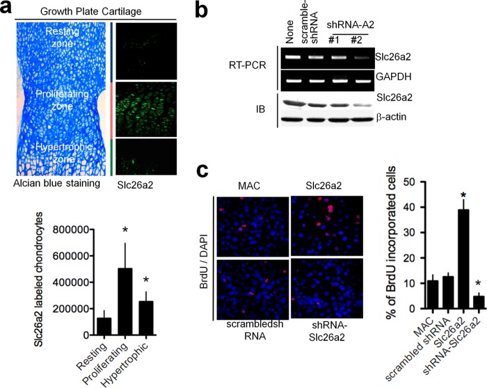
Slc26a2 stimulates chondrocyte proliferation. a, localization of Slc26a2 in a 5-day-old mouse epiphyseal growth plate. Growth plate zones were identified by Alcian blue staining, and the number of chondrocytes expressing Slc26a2 in each zone was determined using ImageJ software (columns). The averages are the mean ± S.E. of four experiments. b, shRNA #2 reduced the expression of Slc26a2 mRNA and protein in MACs by about 70% after 72 h of treatment. IB, immunoblot. c, expression of Slc26a2 increased, whereas knockdown of Slc26a2 decreased, proliferation of MACs. Cells were treated with Slc26a2 and shSlc26a2 for 2 days before an assay of cell proliferation by BrdU staining (n = 3). *, p < 0.05.

