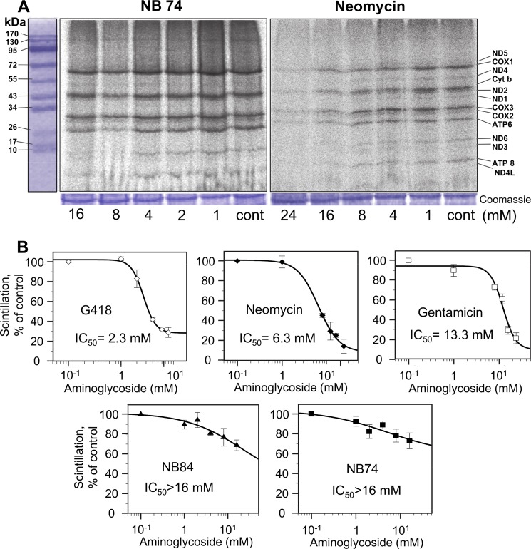
FIGURE 2.
Dose-response effects of AGs on mitochondrial protein synthesis as determined in whole cell assay. HeLa cells were incubated with varied concentrations of G418, neomycin, gentamicin, NB74, and NB84 as indicated. A, after lyses, equal amounts of protein (Coomassie staining) were fractionated on SDS-PAGE, and [35S]methionine-labeled mitochondrial protein levels were determined by densitometry. Tentative identities of labeled mitochondrial proteins were determined as reported (23): COX1 (57 kDa), COX2 (26 kDa), COX3 (29 kDa), subunits I, II, and III of cytochrome c oxidase; cytochrome b (44 kDa) apocytochrome b of the bc1 complex; ATPase 6 (25 kDa), and ATPase 8 (10 kDa) subunits 6 and 8 of the F0 portion of ATP synthase; ND1 (35 kDa), ND2 (39 kDa), ND3 (13 kDa), ND4 (52 kDa), ND5 (67 kDa), ND6 (18 kDa), and ND4L (8 kDa), subunits of NADH dehydrogenase. B, semilogarithmic plots of scintillation counting as a function of AG concentration. Radioactivity was measured on the lysed mixture after acid precipitation as described under “Experimental Procedures.” The 50% inhibitory concentrations (IC50Mit) were determined by Grafit5 software. The data represent means ± S.D., n = 3/point.

