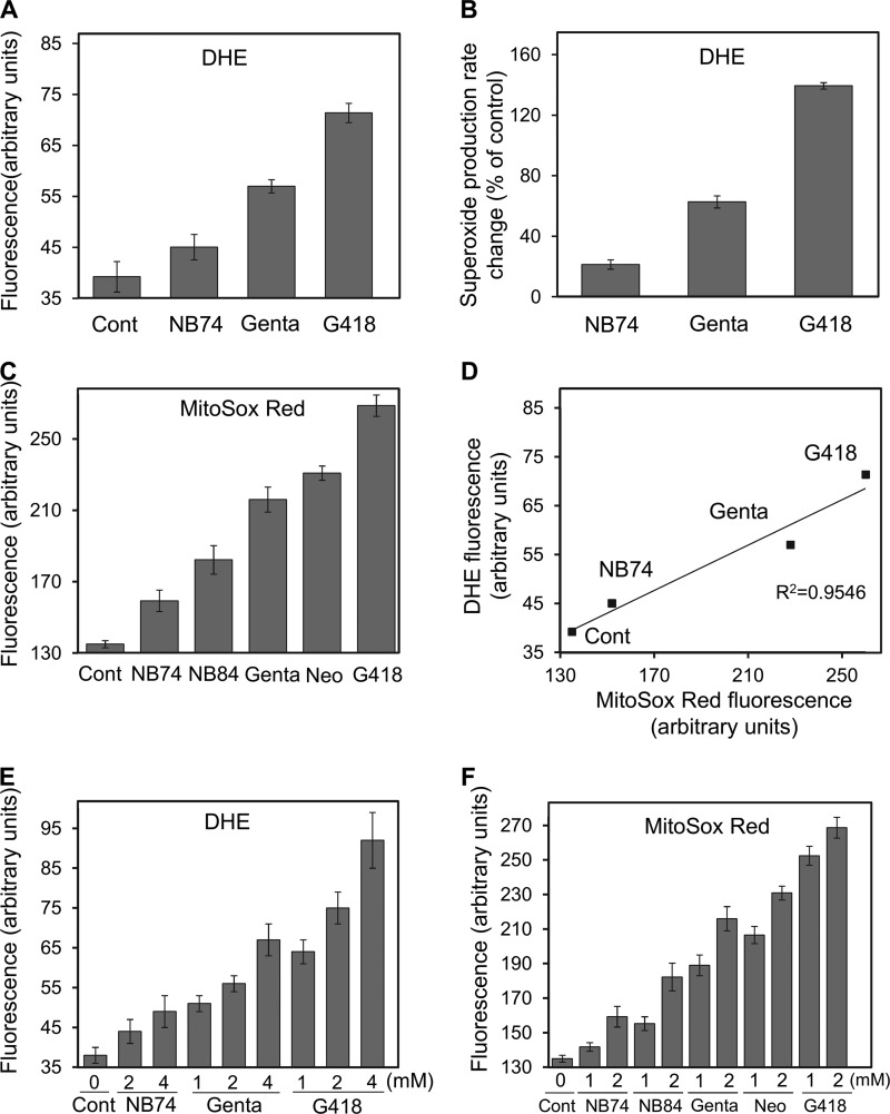
FIGURE 4.
Effect of AGs on superoxide radical production. HeLa cells were incubated with a series of AGs as indicated, and the superoxide radical formation was detected by using two different fluorescence probes. A, DHE (1 μm) probe, measuring fluorescence at 518/605 nm. The data represent means ± S.D., n = 3/point. B, rate changes (as a percentage of control) of superoxide radical production under DHE treatment. The data represent means ± S.D., n = 3/point. C, MitoSox Red (1 μm) probe, measuring fluorescence at 510/580 nm. The data represent means ± S.D., n = 3/point. D, plot of the superoxide radical levels as measured by DHE fluorescence (data from A) against the levels obtained from MitoSox Red (data from C) (linearity R2 = 0.9546). E and F, dose-dependent effect of AGs on superoxide production. Following the treatment with AG (24 h), cells were incubated with DHE (1 μm, E) or MitoSox Red (1 μm, F) for an additional 170 min, 37 °C, and the fluorescence was measured as above. The data represent means ± S.D., n = 3/point. Cont, control; Gent, gentamicin; Neo, neomycin.

