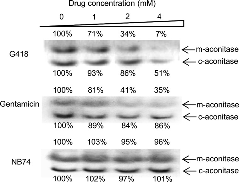
FIGURE 5.
In-gel activity assay of aconitase enzyme from mitochondrial and cytoplasmic origin (m-aconitase and c-aconitase), in HeLa cells Triton lysate as a function of AG concentration as indicated. HeLa cells were incubated with different AGs for 24 h. After lysis, the lysates (5 mg protein/ml) were subjected to electrophoresis, followed by activity quantization (29) as described under “Experimental Procedures.”

