FIGURE 7.
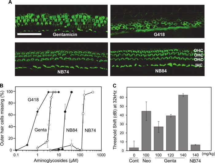
Aminoglycoside-induced hair cell loss in cochlear explants and loss of auditory function in vivo. A, explants of the mouse organ of Corti were incubated for 72 h with a different AGs (as indicated) at concentrations of 3.3 μm and stained for actin as described under “Experimental Procedures.” Following treatment with NB84 and NB74, morphology remains essentially normal, gentamicin causes considerable destruction of hair cells, and cell loss is almost complete with G418. Sections of the basal part are shown. OHC, outer hair cells; IHC, inner hair cells. Calibration bar, 25 μm. B, dose-response curves of missing outer hair cells as a function of AG concentration. Hair cell loss was quantified along the entire length of cochlear explants, and the concentration at 50% loss of hair cells (LC50Coch) was determined by Grafit5 software. The data represent means ± S.E., n = 3–8/point. C, effect of chronic AG treatment (once daily injections at indicated concentrations for 14 days) in vivo on auditory thresholds. Threshold shift is the difference in threshold before and 3 weeks after the end of treatment. The data represent means ± S.D., n = 3/point. Note that dB is a logarithmic scale, i.e., a 10-dB difference indicates a 1 log10 difference in sound intensity. Cont, control; Gent, gentamicin; Neo, neomycin.

