FIGURE 8.

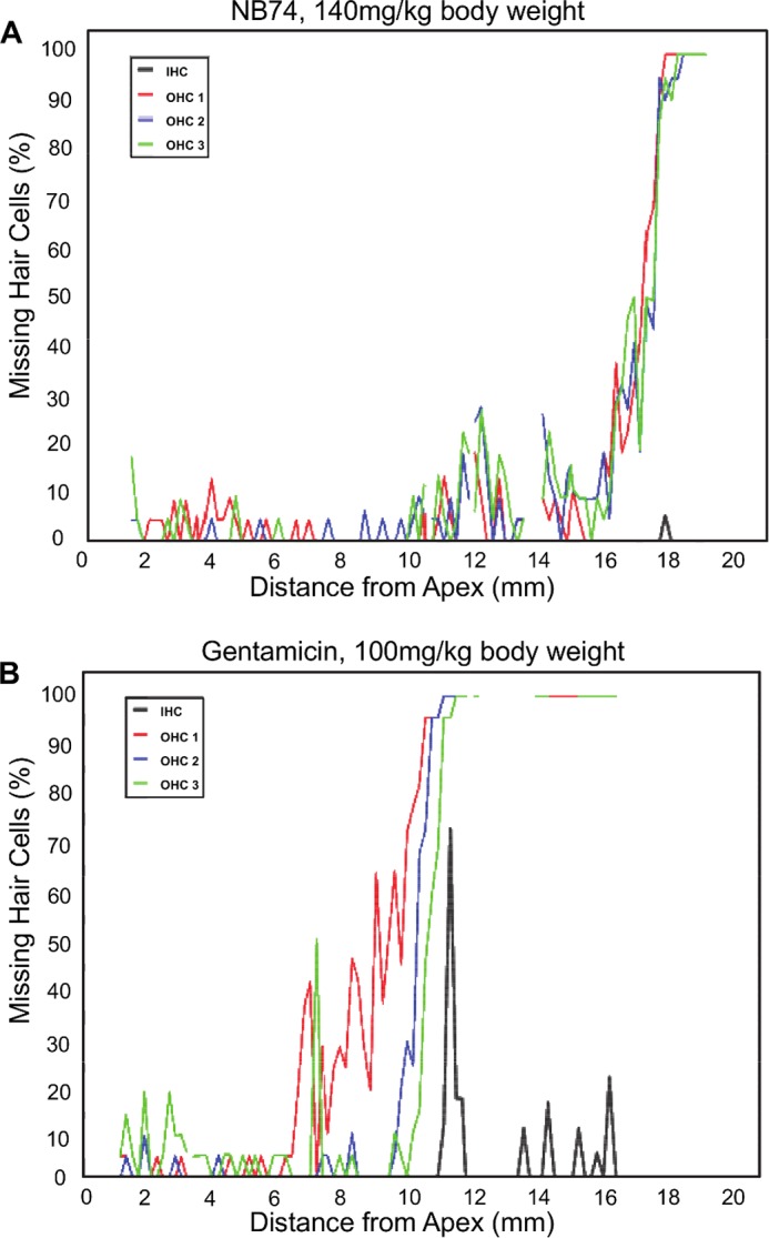
Cytocochleograms showing quantitative evaluation of hair cell loss. Surface preparations of guinea pig cochleae were evaluated quantitatively by counting the presence or absence of hair cells along the entire length of the cochlea. A, after treatment of guinea pigs with NB74 (140 mg/kg of body weight for 14 days), only minimal loss of outer hair cells is observed at the extreme base of the cochlea. B, after treatment with gentamicin (100 mg/kg of body weight for 14 days), loss of outer hair cells (OHC) already begins in the upper middle turn of the cochlea and increases steeply with a total loss in the lower middle and basal turns of the cochlea. Inner hair cells (IHC) show scattered loss toward the base. Representative examples of treatment with gentamicin and NB74 are shown.
